Reversible Injury
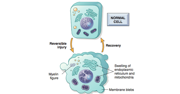
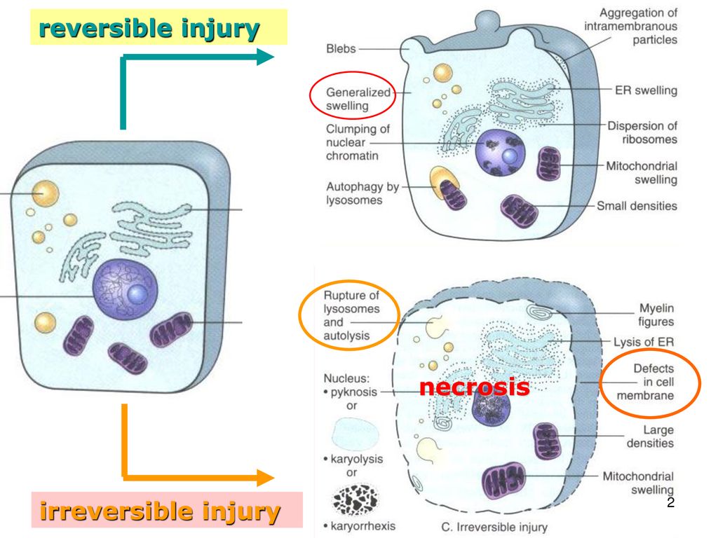
“Cell injury is classified as reversible if the injured cell can regain homeostasis and return to a morphologically (and functionally) normal state”.
- Acute cell swelling is the classic morphologic change in reversible injury.
- Intracellular changes associated with reversible injury includes:
-Plasma membrane alteration i.e. bulging, blunting, loosening of intracellular attachment.
-Mitochondrial changes i.e. swelling.
-Dilation of ER with detachment of ribosomes.
-Nuclear alterations with clumping of chromatin.
Features of Reversible Injury:
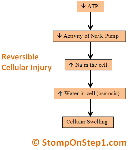
1. Cellular swelling:
- “Cellular swelling or cloudy swelling or hydropic change is characterized by the presence of large vacuoles in the cytoplasm”.
- First manifestation of all form of injury to cells.
- When it affects many cells in an organ, it causes some pallor (as a result of compression of capillaries), increased turgor, increase in weight of the organ.
Microscopic examination:
- Small, clear vacuoles within cytoplasm.
- Cytoplasm shows;
- Swelling of ER, mitochondria.
- Swelling of whole cell.
Mechanism:
- “Failure of Na-K-ATPase pump”
- Failure of cell membrane pump which maintains Na, K balance across the cell membrane.
- Failure of this Na-K-ATPase pump due to def of ATP results in acc. of Na inside and K outside the cell.
- Excess Na inside the cell exerts osmotic pressure and pulls the water inside, resulting in cellular swelling.
- Ribosomes become detached from the RER due to its swelling and therefore protein synthesis reduced.
2. Fatty change:
- “Fatty change is characterized by appearance of lipid vacuoles in the cytoplasm”.
- Principally seen in cells participating in fat metabolism (e.g. hepatocytes, myocardial cells).
- Injured cells may also show increased eosinophilic staining.
Irreversible Injury
“Cell injury is classified as irreversible if the injured cell/cell injury leads to adaptation of the cells and tissue”.
Features of Irreversible Injury:
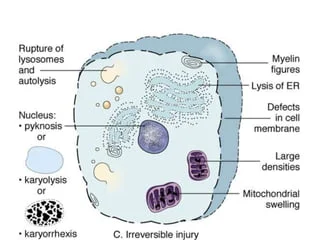
1. Cytoplasmic changes:
- Cells show increased eosinophilia (i.e. pink staining from eosin dye).
- Cells may have a more glassy appearance, homogenous appearance, mostly b/c of loss of glycogen particles.
- Myelin figures; more prominent.
- Mouth eaten appearance; when enzymes have digested cytoplasmic organelles, the cytoplasm becomes vacuolated and appears “mouth-eaten”.
- Irreversible damage to mitochondria manifested by severe vacuolization.
- Extensive damage to plasma membrane.
- Massive calcium influx acting as poison for mitochondria.
- Loss of enzymes and proteins due to increased membrane permeability.
- Lysosomal swelling and leakage of enzymes.
- Electron microscopy shows:
-Discontinuities in plasma and organelle membrane.
-Marked dilation of mitochondria with the appearance of large amorphous densities.
-Intracytoplasmic myelin figures
2. Nuclear changes: (all due to breakdown of DNA and chromatin)
- Karyolysis:
-Basophilia of chromatin may fade.
- Pyknosis:
–Nuclear shrinkage & increased basophilia.
-DNA condenses into a solid shrunken mass.
- Karyorrhexis:
-Pyknotic nucleus undergo fragmentation.
-In 1 to 2 days, nucleus in a dead cell may completely disappear.
-Electron microscopy shows nuclear dissolution.
Leave a Reply